
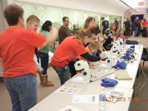
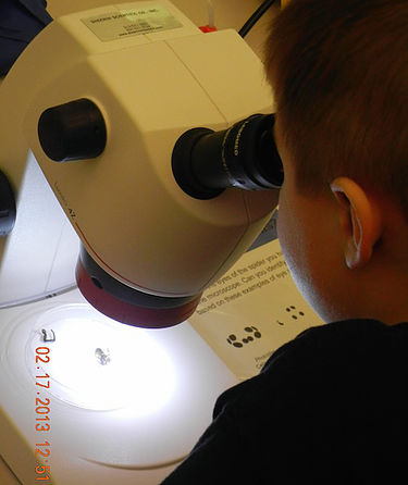
Microscope Madness!
GOAL: It is not very often that people get the opportunity to take a close look at animal body parts; especially body parts of relatively small animals that cannot easily be seen with the naked eye. This station provides an opportunity for participants to see how variable spiders can be. It invites them to look for similarities and differences among different groups of spiders and to think critically and creatively about what this variation might mean in terms of an animal’s lifestyle, habitat, evolutionary history, etc. It also highlights differences between female and male spiders.
BACKGROUND: Although most spiders do not rely heavily upon vision for gathering information from their environment, many spiders non-the-less possess eight eyes. Others, however, have two, or four, or six eyes. Spiders in some families have a single pair of eyes that are much larger than the other pairs – what might this tell you about how important vision is for these animals?
In terms of other sensory systems, spiders are excellent at detecting vibrations that travel through substrates, or surfaces upon which they rest (e.g. webs, leaves, etc.). They use specialized sensory structures called slit sensilla or lyriform organs to detect these vibrations. These structures are best observed under high powered magnification such as a scanning electron microscope (SEM). However, tricobothria, or air-particle movement detectors, can be seen under a dissecting scope. Tricobothria are extremely long, thin hairs that are very sensitive to air particle movements and are thus incredibly important in prey capture. Spiders also rely heavily on chemical information from their environment. They can detect chemical cues through sensory hairs located at various places on their body. Sensory hairs that “taste” chemicals on a surface typically have a single pore at the very tip. On what body parts might you expect chemosensory hairs to be common?
Spiders also have specialized structures for spinning silk (spinnerets), combing out silk (calimistrum), and capturing and immobilizing their prey (fangs with venom glands). Many of these structures are readily seen under a dissecting scope.
PROCEDURE:
-
At each microscope have a bundle of color-coded vials that represent good specimens to examine for each of the observation sheets provided. Download the observation sheets here: eye arrangements, crazy structures, male or female
-
Ideally, have other pictures available to show lyriform organs, contact chemosensory hairs, tricobothria, etc.
-
Allow the participants to get out the specimens and manipulate them under the microscope.

MATERIALS:
-
Dissecting microscopes with lights
-
Petri dishes to hold specimens
-
Squeeze bottles of alcohol
-
Soft forceps
-
Preserved specimens

